 Image credit: Unsplash
Abstract
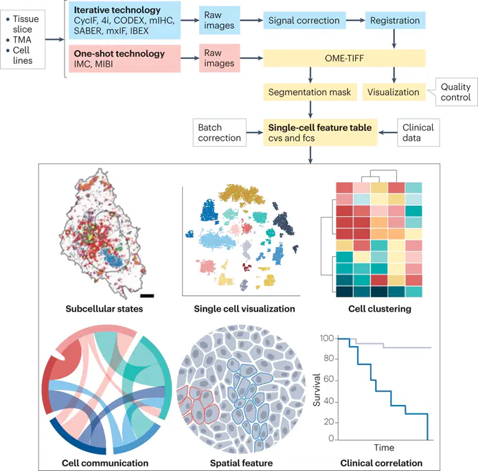
Tissue imaging has become much more colourful in the past decade. Advances in both experimental and analytical methods now make it possible to image protein markers in tissue samples in high multiplex. The ability to routinely image 40–50 markers simultaneously, at single-cell or subcellular resolution, has opened up new vistas in the study of tumour biology. Cellular phenotypes, interaction, communication and spatial organization have become amenable to molecular-level analysis, and application to patient cohorts has identified clinically relevant cellular and tissue features in several cancer types. Here, we review the use of multiplex protein imaging methods to study tumour biology, discuss ongoing attempts to combine these approaches with other forms of spatial omics, and highlight challenges in the field.
This work is driven by the results in my previous paper on LLMs.
Add the publication’s full text or supplementary notes here. You can use rich formatting such as including code, math, and images.
Shan Zhao
Principal Investigator
My research interests include tissue transparency, spatial omics and artificial intelligence.